
Figure 4.
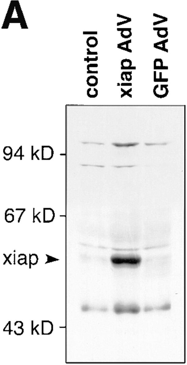


Inhibition of apoptosis by ectopic xiap expression. (A) Lysates of noninfected or infected HUVECs were separated by SDS-PAGE, blotted onto nylon membranes, and stained for myc-tagged XIAP protein. AdV, adenovirus; GFP, green fluorescent protein. (B) HUVECs were infected with IκBα alone (c and d), together with xiap (e and f ), or together with GFP (g and h) recombinant adenovirus. 48 h after infection cells were treated with TNF-α (500 U/ml) for 6 h or left untreated and analyzed by FACS® after propidium iodide staining. Cells with a DNA content <2 N appear in the sub-G1 region (M1). The percentage of cells found in the M1 region is indicated. The data show one out of three representative experiments.