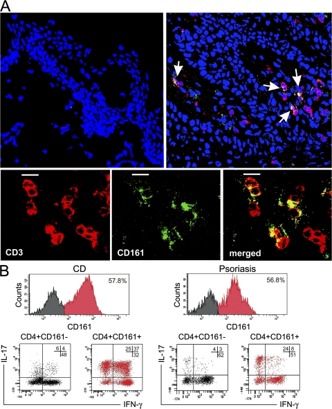
Figure 3.
Detection of CD161+ T cells in inflamed tissues and demonstration of their ability to produce IL-17. (A) Evaluation of the expression of CD3 (red) and CD161 (green) in skin from a healthy donor (top left) or in lesional skin from a psoriatic patient (top right) by confocal microscopy. Arrows point to cells showing double labeling for CD3 and CD161 (yellow) in the skin of the psoriatic patient. TO-PRO-3 (Invitrogen) counterstained nuclei. (bottom) A close-up of CD3+CD161+ double-positive cells (merged; yellow) is shown. Images obtained in the skin of one out of three healthy or psoriatic donors are depicted. Bars, 10 μm. (B) Infiltrating T cells recovered from gut areas of subjects with CD or skin biopsies of subjects with psoriasis were expanded in vitro for 1 wk and assessed for CD161 expression, as well as for their ability to produce IFN-γ and IL-17 after stimulation with PMA plus ionomycin. Representative flow cytometric analysis obtained in one out of three subjects with CD and in one out of three subjects with psoriasis are shown. Percentages of gated cells are shown.

