Figure 6.
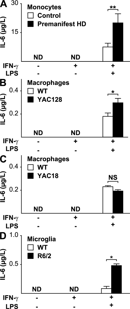
HD monocytes, macrophages, and microglia are overactive when stimulated. (A) No IL-6 was detectable in the supernatant of monocytes from control (n = 9) or premanifest HD subjects (n = 8) in the unstimulated state or after priming with IFN-γ. Monocytes stimulated by the addition of both IFN-γ and 2 μg/ml LPS expressed IL-6, but expression levels were significantly higher from HD monocytes. (B) Alveolar macrophages from the YAC128 HD mouse model have similarly altered function when stimulated. YAC128 macrophages stimulated by the addition of both IFN-γ and 100 ng/ml LPS expressed significantly more IL-6. n = 3 WT and 4 YAC128. (C) Macrophages from YAC18 mice, which differ from YAC128 cells only in the number of CAG repeats, behaved no differently from WT cells (P = 0.231; n = 4 per genotype) in response to stimulation at the same LPS concentration, suggesting that the hyperactivity in the YAC128 is due to mutant huntingtin. (D) Microglia isolated from neonatal R6/2 mice are also hyperactive when stimulated by 10 ng/ml LPS (n = 4 per group). Graphs show mean concentrations with standard error bars. ND, not detected. Unpaired t tests: *, P < 0.05; **, P < 0.01.

