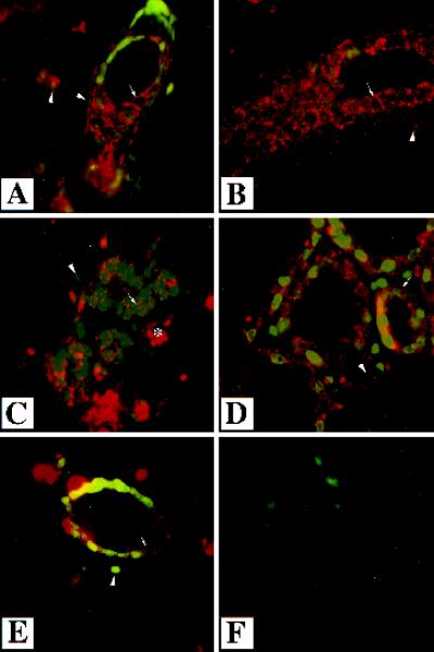
Figure 3.
Localization of endogenous TGF-β type II receptor in the mammary gland. Immunofluorescent analysis was performed using an antibody directed to the TGF-β type II receptor on sections of mammary glands from wild-type mice that were virgin (A and B), 12.5 d pregnant (C), lactating 1 d (D), or involuting 3 d (E). Sections were counterstained with Yo-pro. White arrows indicate staining surrounding epithelial cells and presumably membrane associated. White arrowheads indicate the nuclei of stromal–myoepithelial cells, which also stain for the type II receptor. Immunoreactivity was detected in the stroma and the epithelium in all stages of development. No immunoreactivity was detected in the absence of primary antibody (F). The asterisk denotes a blood vessel. Magnification, 630×.

