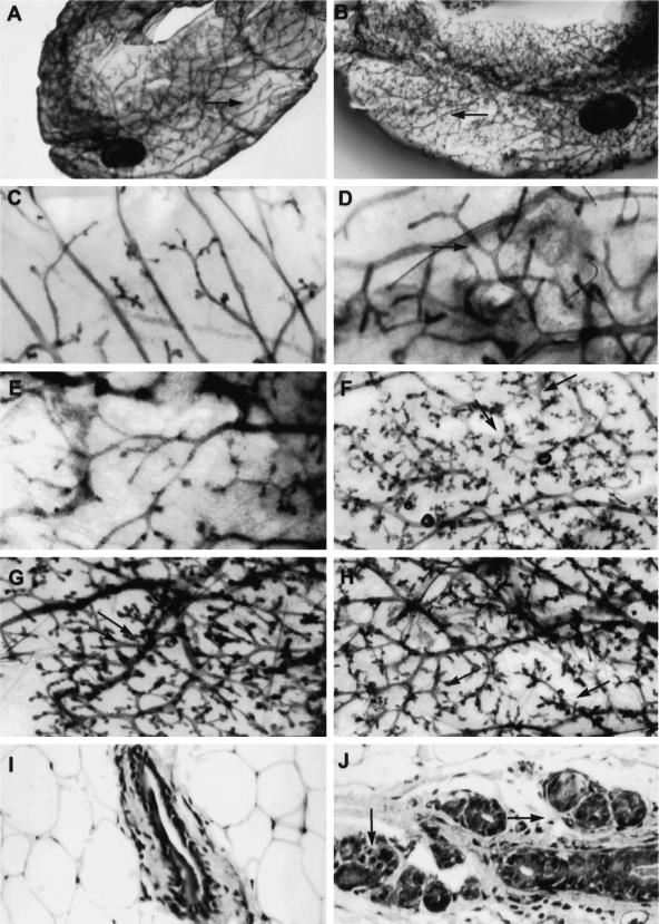
Figure 6.
Morphology and histology of MT-DNIIR mammary glands. (A–H) Hematoxylin-stained whole-mount preparations of untreated wild-type (C) and MT-DNIIR-28 (E) or zinc-treated wild-type (A and D), MT-DNIIR-28 (B and F), MT-DNIIR-4 (G), and MT-DNIIR-27 (H) virgin mammary glands. Note the open spaces along the ducts in the wild-type glands (A and D, arrows), compared with the highly branched structures in the transgenic glands (B, F, G, and H, arrows). (I and J) Hematoxylin and eosin–stained sections of zinc-treated wild-type (I) and MT-DNIIR-28 (J) virgin mammary glands. Note the clusters of small ducts surrounding the larger duct in the transgenic gland (J, arrows) compared with the single duct in the wild-type gland (I). Magnification, A and B, 3.5×; C–H, 15×; I and J, 200×.

