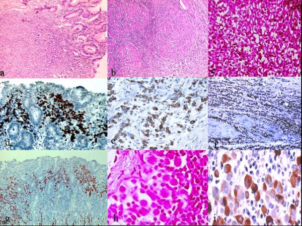
Figure 1.

Photomicrographs of stomach. a)Small monotonous cells arranged in single elements crowed in mucosal layer (Haematoxylin-eosin original magnification 10×). b) Lenities plastic-like invasion of muscular layers (H&E original magnification 20×). c) Neoplastic cells with signet ring-like appearance: presence of an admixture of signet ring cells with single sharply circumscribed vacuoles and multivacuolated forms (hematoxylin-eosin, original magnification 40×). d-e) Neoplastic cells show a strong expression for cytokeratin 7 (original magnification 20×; 40×). f-g) Tumors cells show diffuse and strong nuclear positivity for oestrogenic receptors (original magnification 10×; 20×). h) Focus of Neoplatic cells. i) Cytoplasmatic positivity for gross cystic disease fluid protein 15.
