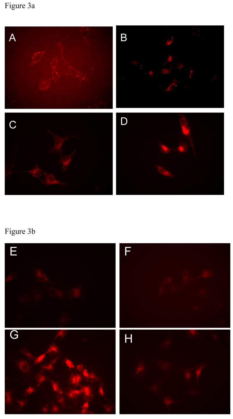
Figure 3.
Figure 3a. A549 cells treated with 1 μM of compounds (A) 5, (B) 6, (C) 9, and (D) 10 for 1 h at 37 °C. Arbitrary intensity scale is 300 to 650 for 5, 6, and 9. The intensity scale for 10 is 300 (black) to 1500 (red) because of the exceptionally high fluorescence intensity relative to other compounds in the figure.
Figure 3b. A549 cells incubated with 1μM of cypate (E and F) and 9 (G and H) for 45 min 37 °C, without (E and G) or with (F and H) 1 h pretreatment with 100 μM of cyclo(RGDfV) that binds ABIR. The intensity scale for 1128 ms exposure is 200 (black) to 600 (red).

