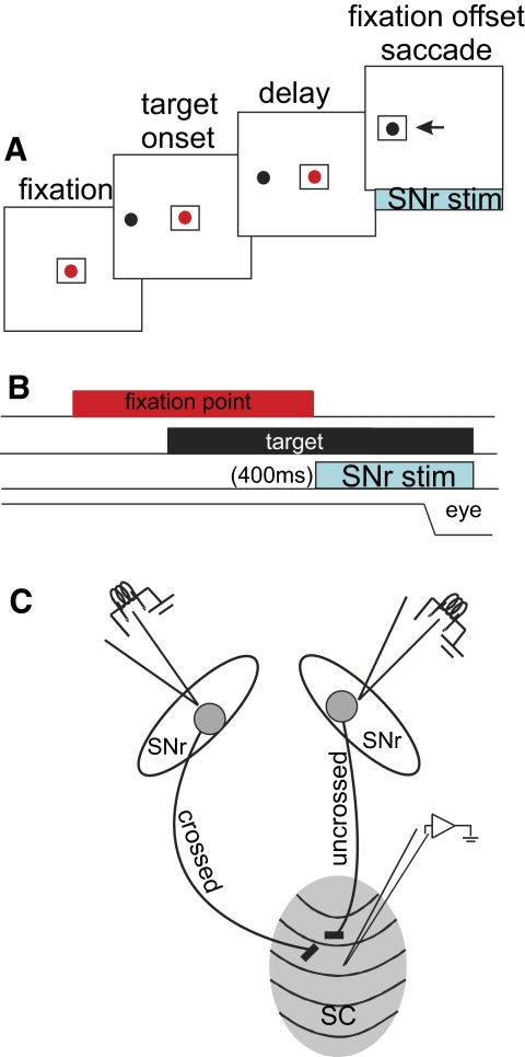
FIG. 1.
Behavioral, stimulation, and recording procedures. A: a schematic depiction of the spatial arrangement of the task. Each square indicates the screen at which the monkeys looked. The red spot is the fixation spot and the black spot is the target. The example shows a visually guided saccade made to the left hemifield. The arrow indicates the saccade. The blue rectangle below the labeled “SNr stim” indicates the part of the task in which stimulation of the substantia nigra pars reticulata (SNr) occurred. This occurred coincident with the fixation point offset. B: the temporal arrangement of the visually guided, delayed-saccade task. The SNr stimulation indicated by the blue rectangle lasted for 400 ms beginning at the onset of the fixation spot offset. The line labeled “eye” is a schematic of the eye position. C: schematic arrangement of the physiological procedures. See methods for details. The ellipses are schematics of left and right SNr nuclei. The gray oval is a schematic of the right superior colliculus (SC). The 2 SNr nuclei were stimulated (independently) while neurons in the SC were recorded independently; “crossed indicates the SC on the side contralateral to the stimulation SNr and “uncrossed” indicates the SC on the side ipsilateral to the stimulation SNr.

