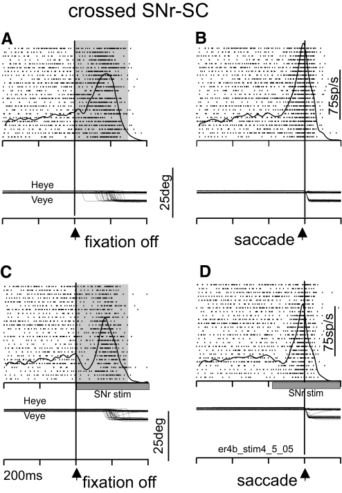
FIG. 4.
SNr stimulation suppresses contralateral SC neuronal activity. The arrangement of this figure is the same as Fig. 3. A and C are aligned on fixation point onset. B and D are aligned on saccade onset. A and B are without SNr stimulation. C and D are with SNr stimulation. Heye, horizontal eye position; Veye, vertical eye position; fpoff, fixation point offset. Each tick on the time axis is separated by 200 ms (er4b_stim4_5_05 is the filename used for reference).

