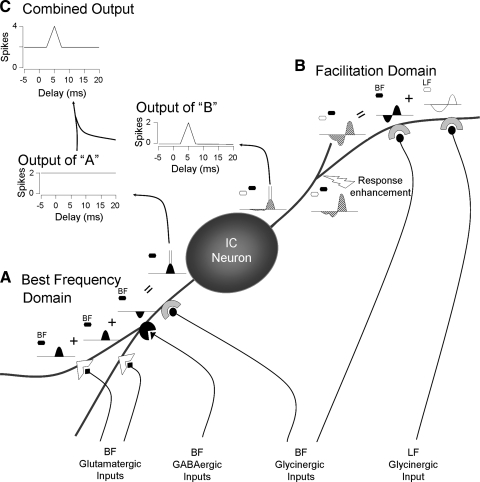
FIG. 13.
Schematic representation of processing domains for an IC neuron. We propose that ascending inputs onto IC neurons are functionally and perhaps anatomically localized. In this figure, sounds (hexagons) and postsynaptic potentials (curved shapes) in response to best frequency (BF) sounds are indicated by filled hexagons and curves, respectively; unfilled shapes refer to lower frequency (LF) sounds and potentials. Hatched potentials result from combinations of BF and LF sounds. A: glutamatergic, GABAergic, and glycinergic inputs tuned near the BF interact among themselves to generate and shape responses near the neuron's BF (bottom left). These inputs generate action potentials that are part of the neuron's output. Since inputs within this domain are affected only by sounds near the BF, spikes generated by interactions within this domain are independent of the delay between a LF sound and a sound near BF. B: the activity within the BF domain does not affect the facilitatory interaction within the facilitation domain (top right). These results and previous work (Sanchez et al. 2008) strongly suggest that 1) the BF and LF glycinergic inputs contributing to facilitation are segregated from other inputs, 2) that they are electrically isolated from the soma, and 3) that an active mechanism (response enhancement) is required to trigger facilitation spikes. Because of selective enhancement of the depolarization and overall decrement caused by cable properties, the voltage changes arriving at the soma and spike generator are sufficient to trigger spikes but separate postsynaptic potentials are too small to be observed. C: the combined output reflects the activity in both domains. Blockade of glycine receptors typically results in a delay function similar to that of the BF domain (Nataraj and Wenstrup 2005; Wenstrup and Leroy 2001). In contrast, blockade of ionotropic glutamate receptors results in a delay function like the output of the facilitation domain (Sanchez et al. 2008). Figure modified from Sanchez et al. (2008).

