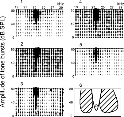
FIG. 5.
Inhibition of a collicular PLS neuron evoked by electric stimulation of BF-matched cortical neurons. The BFs of the recorded collicular and stimulated cortical neurons were the same, 23.5 kHz. The frequency-amplitude (F-A) scan repeated 5 times. The size of a dot is proportional to the number of spikes/tone burst. 1: control, i.e., F-A scan only. 2: F-A scan with a probe tone at 23.5 kHz and 40 dB SPL. The probe tone alone excited the neuron, evoking 0.6 spike/probe on the average. 3: F-A scan with the probe 30 min after the onset of cortical electric stimulation. 4: F-A scan with the probe 60 min after the electric stimulation. 5: F-A scan only 80 min after the electric stimulation (recovery). In 3, the cortical electric stimulation sharpened the excitatory area (excitatory frequency-tuning curve) and produced the large inhibitory areas on both sides of it. In 6, the excitatory and inhibitory areas in 3 are schematically drawn. The open circle indicates the probe tone.

