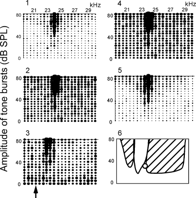
FIG. 7.
Inhibition of a collicular non-PLS neuron evoked by electric stimulation of BF-unmatched cortical neurons. The F-A scan repeated five times as in Fig. 5. 1: control, i.e., F-A scan only (control). 2: F-A scan with a probe tone at 24 kHz and 35 dB SPL. The probe tone alone excited the neuron, evoking 0.8 spikes/probe on the average. 3: F-A scan with the probe 20 min after the onset of the electric stimulation. Stimulated 21.0-kHz-tuned cortical neurons (↑). 4: F-A scan with the probe 60 min after the electric stimulation. 5: F-A scan 80 min after the electric stimulation (recovery). In 3, the BF shifted from 24 to 23 kHz; an inhibitory area was large at the side higher than the excitatory area, but small at the side lower than that. In 6, the excitatory and inhibitory areas in 3 are schematically drawn. ○, the probe tone.

