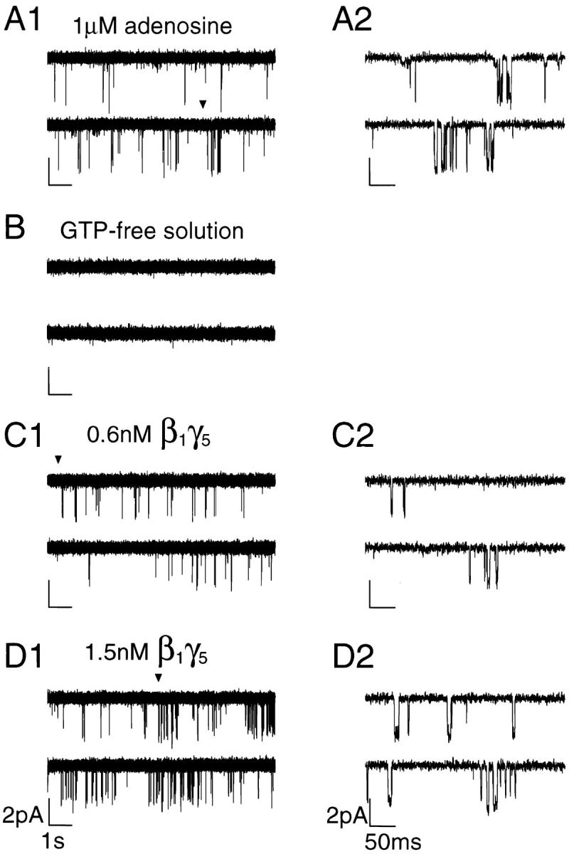
Figure 5.

Activation of the KACh channels by recombinant Gβ1γ5. (A1) Representative single-channel activity recorded in cell-attached configuration from an atrial myocyte in the presence of 1 μM adenosine. The membrane potential was clamped at −90 mV. (A2) Part of the trace in A1 (arrowhead) has been expanded to show the transitions between the closed and open states of the channel with a higher resolution. (B) Channel activity disappeared after the patch excision in GTP-free solution. The membrane potential continued to be clamped at −90 mV for the entire experiment. (C–D) Application of nanomolar concentrations of Gβ1γ5 (0.6 nM in C1 and 1.5 nM in D1) restored the channel activity in a concentration- dependent manner. Extended current traces starting at the points indicated by the arrowheads in C1 and D1 are shown on the right in C2 and D2, respectively.
