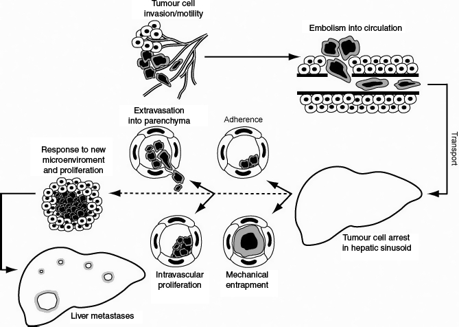
Figure 2.

This figure graphically depicts the metastatic process, focussing on the early stages. Initially the colorectal cancer cells at the primary locus proliferate. Subsequently, these malignant cells become invasive and adopt a migratory phenotype. The invasion process if successful will allow the tumour cells to enter the circulation. In vivo, colorectal cancer cells can metastasize using a haematogenous or lymphatic method. For simplicity, only haematogenous spread is discussed. The haematogenous route of spread is utilized in many recent in vivo studies shown in Table 1. The cancer cells are then carried via the portal circulation to the liver where the sinusoids are the vessels with the smallest diameter. The figure then depicts the two different methods proposed for tumour cell arrest – mechanical entrapment and specific tumour cell adhesion. After the initial arrest, there is also evidence to support different methods of early metastatic development. Tumour cells can either undergo extravasation into the parenchyma and subsequent proliferation or undergo initial proliferation intravascularly followed by liver infiltration. If the tumour cells successfully complete these stages, they proliferate within their new environment and can develop into macroscopic liver metastasis.
