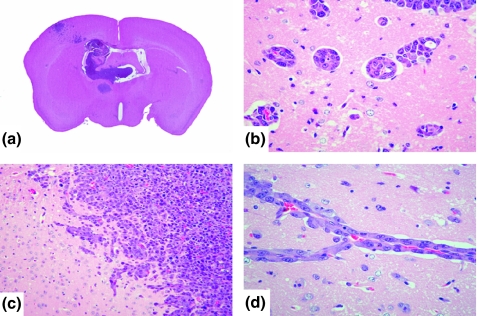
Figure 1.
(a) Macroscopic findings (H&E staining) of brain metastasis of internal carotid artery injection. Tumour foci of brain metastasis are present in the posterior limbic area and Ammon’s horn, in addition to the agranular motor cortical area and somatosensory cortex. (b, d) The metastatic foci were localized in the perivascular lesion. (c) Invasive proliferations, neoplastic cells infiltrated the brain parenchyma, the border of which had become indistinct.

