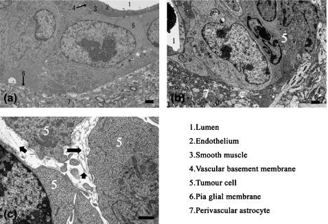
Figure 5.
Electron microscopic observation. (a) Perivascular proliferation. Neoplastic cells were localized between the vascular basement membrane and pia-glial membrane (b) Invasive proliferation. Neoplastic cells were observed in the brain parenchyma, and the pia-glial membrane was absent. (c) Fragments of collagen fibres were observed in the gap between neoplastic cells. Arrows point to the fragment of collagen fibres.

