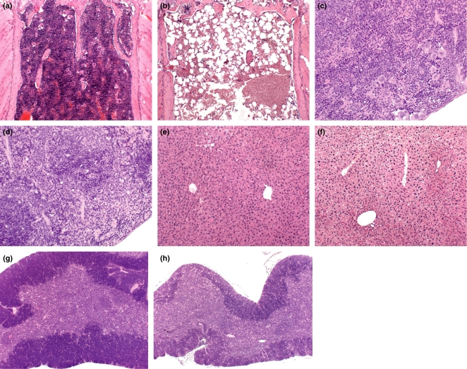
Figure 6.
Haematoxylin and eosin stained sections of sternum, spleen, liver and thymus from control and azathioprine- (AZA-) treated mice (100 mg/kg daily for 10 days). (a) Sternum from a control (vehicle-treated) mouse at day 1 postdosing showing normal marrow cellularity. [Original magnification (OM) × 100.] (b) Sternum from an AZA-treated mouse at day 1 after the final dose showing significant depletion in the cellularity of the marrow, with increased numbers of adipocytes. (OM × 100.) (c) Spleen from a control (vehicle-treated) mouse at day 9 postdosing, showing the normal appearance of the spleen. (OM × 200.) (d) Spleen from an AZA-treated mouse at day 9 postdosing showing the absence of extramedullary haemopoiesis. (OM × 200.) (e) Liver from a control (vehicle-treated) mouse at day 1 postdosing, showing the normal appearance of the hepatic lobules. (OM × 100.) (f) Liver from an AZA-treated mouse at day 1 postdosing showing centrilobular hypertrophy; hepatocytes show cytoplasmic vacuolation consistent with glycogen vacuolation/rarefaction. (OM × 100.) (g) Thymus from a control (vehicle-treated) mouse at day 9 postdosing. (OM × 50.) (h) Thymus from an AZA-treated mouse on day 9 postdosing showing marked atrophy. (OM × 50.)

