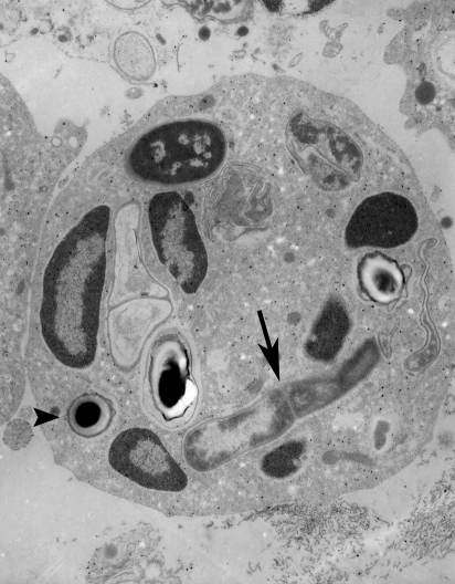
Figure 3.
Bacillus anthracis organisms at 6 h after inoculation of spores onto abraded skin of an HRS/J hr/hr mouse. Electron micrograph was taken of an epon section made from the filter onto which the spores were placed during inoculation. Note that ungerminated spores (arrowhead) and vegetative bacilli (arrow) have been ingested by a neutrophil (original magnification of 8000×).

