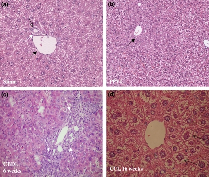
Figure 2.
Haematoxylin–eosin staining. Normal liver histology is seen in sham-operated (a) (magnification ×20) and PPVL (b) (magnification ×10) mice. A portal tract is shown consisting of a portal vein (black arrow), bile duct (white arrow) and hepatic arteriole (open arrow). Marked proliferation of bile ductules is seen in CBDL mice after 6 weeks of induction (white arrows, c) (magnification ×20). Hepatocytes are swollen (arrows) and surrounded by fibrotic tissue with a tendency of dissection of groups of hepatocytes after a longer period of CCl4 induction (d) (magnification ×20). PPVL, partial portal vein ligation; CBDL, common bile duct ligation; CCl4, carbon tetrachloride.

