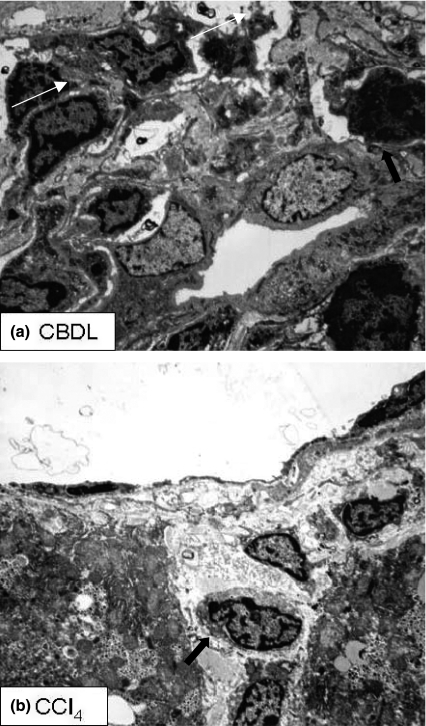
Figure 5.
Electron microscopy. (a) Portal tract of CBDL mice (4 weeks). Proliferating bile ducts are surrounded by inflammatory cells (arrows). (b) Central vein area of CCl4 mice (8 weeks). Degenerating hepatocytes undergo necrotic changes (arrows) and are surrounded by inflammatory cells. CBDL, common bile duct ligation; CCl4, carbon tetrachloride.

