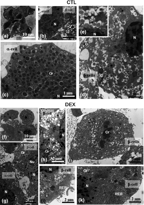
Figure 4.
Transmission Electron Microscopy of isolated islets. General view of the islets (a, f); partial view of the cytoplasm of the alpha and beta cells showing the characteristic granules in CTL and DEX group (b, g, respectively); detailed view of the alpha-cell cytoplasm (c) and of the beta-cell cytoplasm in the CTL group (d) with details of granules (inset). Note the increase in the secreting organelles, such as the Golgi apparatus, in the beta-cell cytoplasm (h) and rough endoplasmic reticulum in the peripheral region (i, k) in DEX islet. Mitosis (initial telophasis) figure in the beta cell of DEX islet (j). N, nucleus; Gr, granules; C, centrioles; Cr, chromosomes; Nu, nucleolus; GA, Golgi apparatus; RER, rough endoplasmic reticulum.

