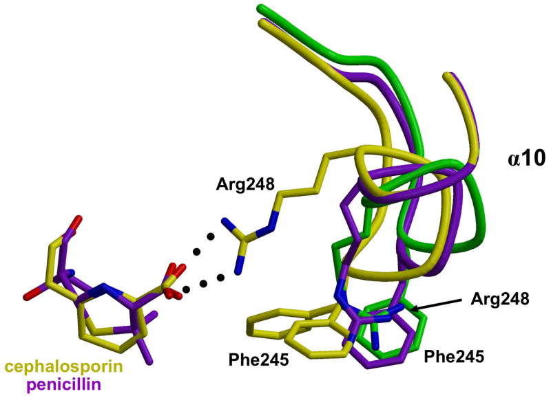
Figure 2.
Interaction between Arg 248 and the cephalosporin carboxylate. The backbones of wild-type (green), penicillin 5-bound (purple) and cephalosporin 6-bound (yellow) structures of PBP5 are superimposed showing conformational differences in a loop comprising residues 242–248. In the cephalosporin structure, Arg 248 interacts with the carboxylate of the cephalosporin (colored yellow) whereas in the penicillin-bound structure Arg 248 occupies a position similar to that in wild-type PBP5. There are also differences in the respective positions of Phe 245. Figure prepared using MOLSCRIPT53 and RASTER3D.54

