Figure 4.
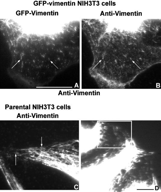
Movie 4: Fountains—the movements of IF fragments. IF fragments are present in GFP-vimentin–expressing cells and in the parental NIH3T3 cells. (A and B) GFP-vimentin–expressing NIH3T3 cells. (A) GFP fluorescence. (B) Indirect immunofluorescent pattern of the same cells fixed and stained as described by Gurland and Gundersen (1995) using a polyclonal antibody (1274) (Wang et al., 1984). Arrows point to two of the many fragments present. (C) NIH3T3 cells fixed and stained as in B. Arrows point to IF fragments present in the parental cells. (D) First frame of a movie in which the IF fragments display dynamic movements. The movie is from the region enclosed in the box and is played four times. There is a pause only after the first time through the movie. Bars: (A, B, and D) 10 μm; (C) 5 μm.

