
Fig. 7.
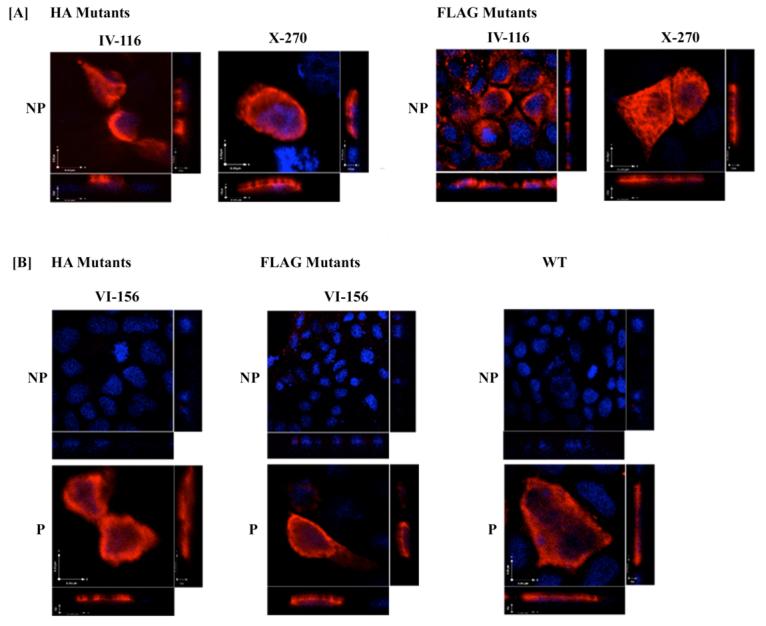
Cellular localization of epitope-tagged mutants in polarized MDCK-II cells. MDCK-II cells, grown on collagen-treated chambers slides, were transiently transfected with native hASBT (WT) and mutant hASBT constructs. 72 hours post transfection, cells were processed for immunofluoresence confocal laser scanning microscopy under permeabilizing (P) and nonpermeabilizing conditions (NP). Panel A displays cells expressing mutants that localize on the extracellular plasma surface, whereas panel B represents cells that express epitope constructs that can only be visualized after saponin treatment (P), indicative of intracellular localization. Native hASBT was used as a positive control to depict intracellular and apical localization. Images represent orthogonal 3-D profiles with the inset view defining the XY axis and the outer panels reveal the YZ (right side) and XZ (bottom side) focal planes. Epitope-tagged mutants and native hASBT exhibited apical localization (red fluorescent signal) relative to the DAPI-stained cell nucleus (blue signal). Fluorescence signal was absent at the basolateral side. All stacked images were acquired using 512×512 pixel resolution under a 60× oil objective. All images were subjected to iterative deconvolution set at 99% confidence using the calculated point spread function with the Volocity 3.6 software. Scale bar, 10 μm.