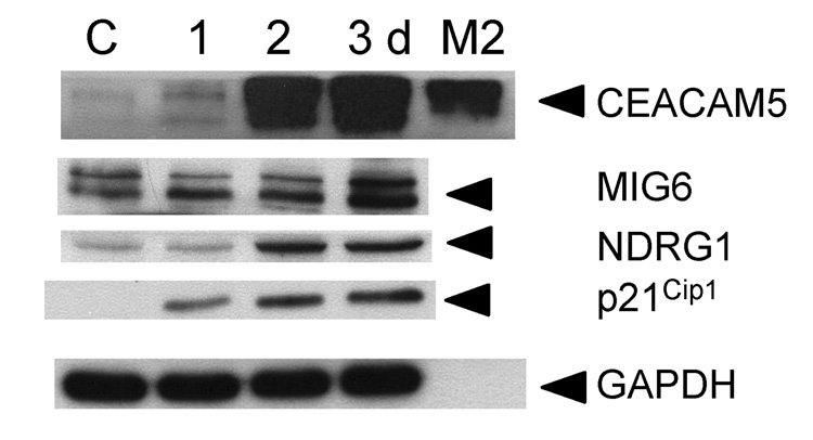
Figure 2. Expression of selected differentiation markers in perifosine treated PC-3 cells.
PC-3 cells were treated with 5 µM perifosine for indicated periods of time. Attached cells were harvested, washed in PBS, and lysed as described in Material and Methods. Conditioned media (M2) was collected and concentrated to analyze secretion of CEACAM5 two days after perifosine treatment. Total proteins at 30 µg were separated in a 10% polyacrylamide gel and transferred to a PVDF membrane. Membranes were incubated with an anti-CEACAM5, MIG6, NDRG1, and p21Cip1 antibodies. Appropriate secondary antibodies labeled with horse peroxidase were used. GAPDH was used to monitor equal protein loading. Membranes were developed as described in Materials and Methods. Experiments were repeated three times and representative blots are presented.

