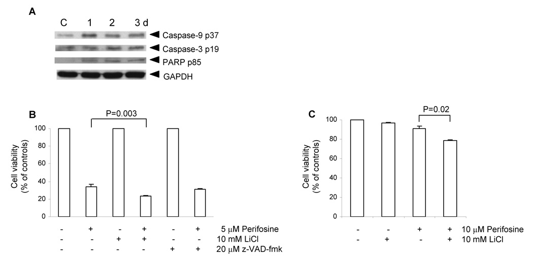
Figure 4. Expression of apoptotic markers by differentiating cells (A), cell viability of PC-3 cells treated with perifosine together with GSK-3β LiCl or pan-caspase z-VAD-fmk inhibitors (B), and viability of LNCaP cells treated with perifosine together with GSK-3β inhibitor LiCl (C).
A, Cell lysates prepared as described in Figure 2 were used to detect protein levels of typical apoptotic markers, such as cleaved caspase-9 p37, caspase-3 p17, and PARP p85 in PC-3 cells treated with 5 µM perifosine for indicated periods of time by means of western blotting as described in Materials and Methods. GAPDH was used to detect equal protein loading, C, control. Experiments were repeated three times and typical results are presented. B, PC-3 cells were seeded at 3 × 10⁵ per well in 6-well plate in triplicates and pre-treated with 10 mM lithium chloride or 20 µM z-VAD-fmk for 1 h prior perifosine treatment at 5 µM for 2 days. Floating and attached cells were collected at the end of treatment, stained with calcein-AM and analyzed by flow cytometry as described in Material and Methods. Bars, SD. C, LNCaP cells were seeded at 2 × 10⁵ per well in 6-well plate in triplicates and pre-treated with lithium chloride for 1 h prior treatment with 10 µM perifosine for 3 days. Floating and attached cells were collected at the end of treatment, stained with calcein-AM and analyzed by flow cytometry as described in Material and Methods. Bars, SD.

