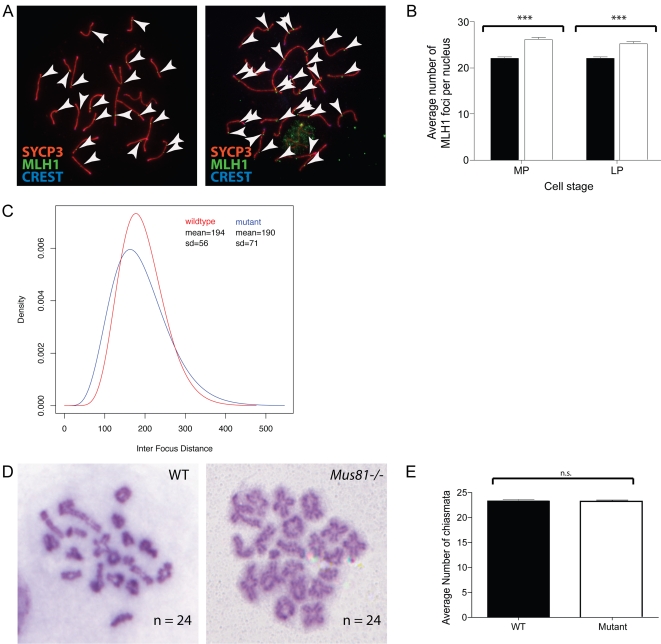
Figure 4. MLH1 accumulation is upregulated in Mus81 nulls, while interference is reduced.
A, B) MLH1 focus numbers are increased in Mus81−/− spermatocytes compared with WT. A) Chromosome spreads from WT (left panel) and Mus81−/− (right panel) spermatocyte stained with antibodies against SYCP3 (red), MLH1 (green) and CREST autoimmune serum (blue). White arrows show the positions of MLH1 foci for clarity. B) Average MLH1 focus numbers counted in mid pachynema (MP) and late pachynema (LP). Statistically significant increases in Mus81−/− counts (white) compared with WT (black) are shown by the asterisks. C) Interference is reduced in the mutant (blue) compared to the WT (red). D, E) Chiasmata counts on cells at diakinesis of prophase I for WT (left) and Mus81−/− (right) mice, chiasmata numbers for each cell are shown, as well as average counts for WT and Mus81−/−.

