Figure 3.
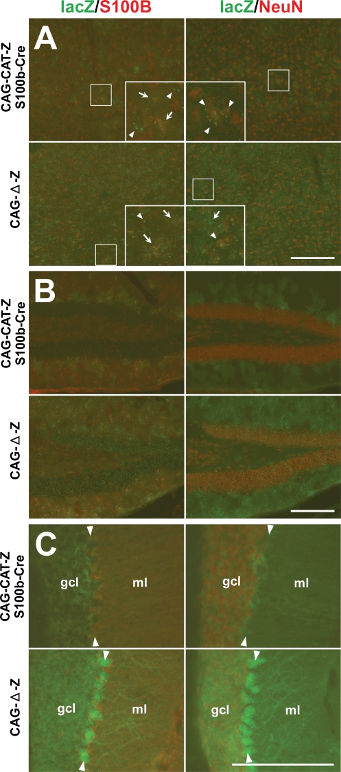
Cell-type specificity of S100b-Cre-mediated recombination in CAG-CAT-Z:S100b-Cre mice. (A–C) Double-immunofluorescence analysis of parasagittal sections from CAG-CAT-Z:S100b-Cre and CAG-Δ-Z (positive control) mice using antibodies directed to β-galactosidase (lacZ), S100B, and NeuN. Left column shows double-labeling of lacZ (green) and S100B (red), and right column shows double-labeling of lacZ (green) and NeuN (red). (A) In the cerebral cortex, lacZ immunoreactivity detected as granules (arrowheads in inserts) was localized in neurons (S100B negative and NeuN positive) in CAG-CAT-Z:S100b-Cre mice, which corresponds to the intensive lacZ staining forming a layer-like pattern in Figure 2A. In contrast, weak and/or unclear lacZ immunoreactivity (arrows in inserts) localizes in astrocytes (S100B-positive and NeuN-negative). LacZ localization both in neurons and astrocytes is less frequent in CAG-CAT-Z:S100b-Cre mice compared to that in CAG-Δ-Z. Each insert is a magnification of the area indicated by the small box in the same panel. (B) LacZ was coexpressed in astrocytes (S100B-positive and NeuN-negative) in the hippocampal dentate gyrus of CAG-CAT-Z:S100b-Cre mice. LacZ/S100B colocalization was less frequent in CAG-CAT-Z:S100b-Cre compared to that in CAG-Δ-Z. (C) LacZ immunoreactivity in the cerebellum of CAG-CAT-Z:S100b-Cre mice localized in astrocytes of the granule cell layer (gcl), Bergmann glial cells in the Purkinje cell layer (arrowheads), and Bergmann glial processes in the molecular layer (ml), all of which were S100B-positive and NeuN-negative. No recombination was detected in the cerebellar granule cells (S100B-negative and NeuN-positive) or Purkinje cells of CAG-CAT-Z:S100b-Cre mice, in contrast to detectable lacZ immunoreactivity in those cells in CAG-Δ-Z mice. Scale bars, 200 μm.

