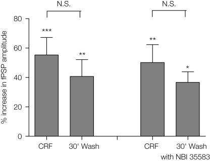
Figure 6.
Comparison of the increase in fPSP amplitude following application of CRF (0.1 μM) and 30 min after wash-out of the peptide in the absence (n = 7 slices from 5 rats) or presence of NBI35583 (1 μM; n = 6 slices from 5 rats). Data shown are the mean ± SEM; *p < 0.05, **p < 0.01, ***p < 0.001, N.S. not significant, one-way repeated measures ANOVA followed by Bonferroni multiple comparison.

