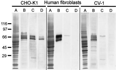
Figure 4.
Characterization of α-B antibody by immunoblotting with total cell homogenates of CHO-K1, human skin fibroblasts, and CV-1 cells. (A) Coomassie R-250 stained gel; (B) immunoblot probed with α-KLC antibody; (C) immunoblot probed with α-B antibody; and (D) immunoblot, probed with α-B* antibody. The positions of the molecular mass markers are shown on the left.

