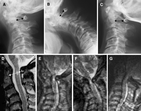
Fig. 1.
Case 1. Dynamic cervical radiographs. In neutral position anterior dislocation of atlas is mild, but the inclination of atlas reduces the spinal canal diameter (a). The anterior dislocation is strongest in flexion (b) and restored in extension (c). Black arrows indicate the anteroposteiror diameter of the bony canal. MRI taken in supine position clearly shows a high intensity lesion in the spinal cord at C1, but does not show the dural/spinal cord compression (d, white arrow). Dynamic MRIs taken in upright position show that the spinal cord at C1 is compressed in any neck position: neutral (e), flexed (f) and extended (g) positions

