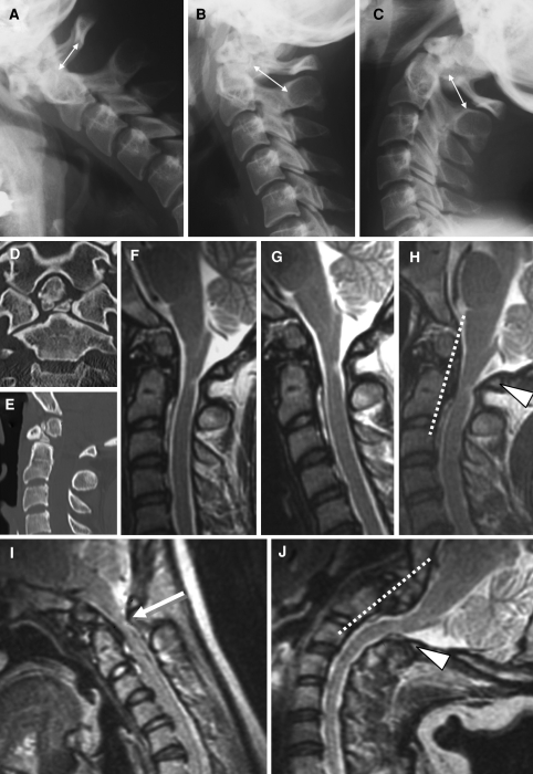
Fig. 2.
Case 2. a–c Dynamic cervical radiographs. The bony canal diameter between the base of dens and the posterior arch of C1 in flexion is lesser than the diameter between dens and C2 lamina in neutral or extension (arrows). d, e Bone-window CT reconstructions show the discontinuity of dens to axis with small bone fragments between them. MRIs taken in supine position show the hyperintensity lesion in the dorsal part of spinal cord at C1 that is compressed in extension (h) but not in neutral (f) nor in flexion (g). Dynamic MRIs taken in upright position show the cord compression in flexion (i, white arrow) but not in extension (j)

