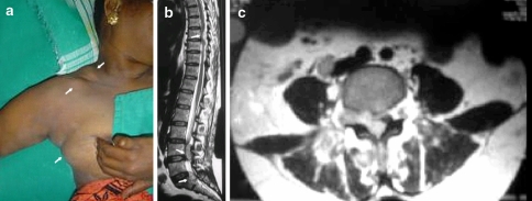
Fig. 1.
a Lymphnodes in the posterior triangle of the neck. Soft tissue swellings in the infra-clavicular region and the right breast. b T2 weighted MRI sagittal section showing the epidural soft tissue lesions at T8, L4 and S1 vertebral levels. c T2 weighted MRI axial section at L4 level demonstrating the epidural mass at the right neural foramina, and lesions in the retroperitoneal and paraspinal region

