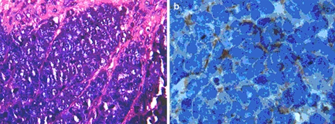
Fig. 2.
a Light microscopy, Haematoxylin-eosin, x400. Demonstrating the typical histology of MCC. Small blue cells with sparse cytoplasm, uniform monotonous medium-sized nuclei and abundant mitoses. Chromatin is displayed in typical salt and pepper pattern. b Immuno-histochemistry staining, x1000, positive for cytokeratin-20 paranuclear dot

