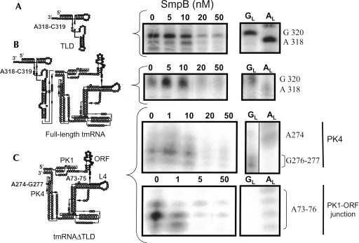
FIGURE 4.
The high-affinity binding site between SmpB and tmRNA is located outside the TLD, as revealed by structural probing. Concentration titration of SmpB with either the TLD (A), tmRNA full length (B), or tmRNAΔTLD (C) revealed by enzymatic structural probes (nuclease S1). Lanes 0, incubation of the RNAs in the absence of SmpB; lanes GL and AL, RNases T1 and U2 hydrolysis ladders, respectively. Accessible nucleotides from either the connecting loop of TLD (gray dots on the TLD structure) or from PK4 in tmRNAΔTLD are protected from S1 cleavages at a 20 nM concentration of SmpB or higher. Nucleotides upstream and at the 3′ end of the internal tag reading frame (gray dots on the tmRNAΔTLD structure) are protected from S1 cleavages at a 1 nM SmpB concentration and higher.

