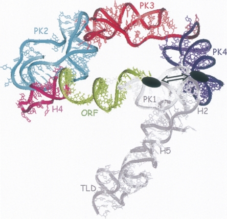
FIGURE 5.
Structure of the minimal tmRNA fragment that contains the higher binding site for the SmpB protein. Reconstruction of tmRNA structure within the electron density derived from the cryo-EM map for the EF-Tu-tmRNA–SmpB complex bound to the ribosome in the absence of ribosomal protein S1 (Gillet et al. 2007). The structural domains of the RNA that are dispensable for the interaction with the protein onto its high-affinity site are colored in gray (PK1, H2, H5, and TLD). The domains that cannot be removed without loosing the binding site are color coded (the ORF is green, H4 is pink, PK2 is blue, PK3 is red, and PK4 is purple). Notice the spatial proximity, emphasized by the double arrow, between the reactivity changes observed upstream of the ORF and in PK4, both marked as black ovals.

