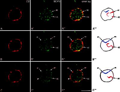
Figure 3.
Double-label immunolocalization with antibodies against lamin C2 (A–C) and protein SCP3 (A′–C′). The labeled rat pachytene spermatocyte was investigated by confocal laser scanning microscopy. Three consecutive optical sections are shown (A–A′, B–B′, C–C′). Overlays are presented in A"–C". A‴–C‴, Schematic representation of the arcs displayed by SCs #1 (blue) and #2 (red). The arrowheads in C′–C" point at an XY body. Bar, 10 μm.

