Figure 4.
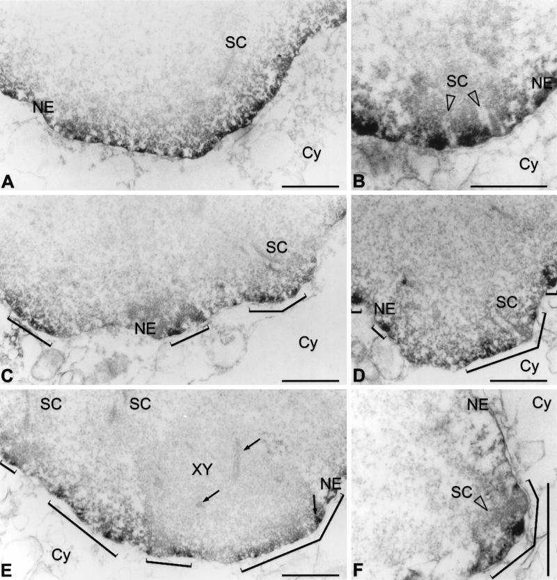
Electron microscopical immunolocalization of lamins B1 (A, B) and C2 (C–F) in pachytene spermatocytes of the rat using peroxidase-conjugated secondary antibodies. The patchy pattern obtained with C2 antibodies is denoted by brackets (C–F). Frontal (B, D) and lateral (F) views of SC attachment sites at the NE are shown. XY, XY body; Cy, cytoplasm. XY body axial elements (including an insertion at the NE) are denoted by arrows (E). Bars, 1 μm.

