Figure 7.
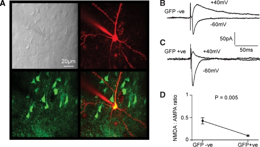
Functional knockout of NMDA-receptor-dependent currents in layer 2/3 pyramidal neurons of fNR1 mice transduced with lentivector encoding GFP and Cre. (A) In vitro brain slices were prepared from fNR1 mice, which had been injected with lentivector encoding GFP and Cre. The brain slice with GFP fluorescence was identified, and a whole-cell recording was made from a GFP-positive layer 2/3 pyramidal neuron imaged using a two-photon microscope. The laser scanned Dodt contrast image (upper left) is aligned with the red fluorescent image of the Alexa Fluor 594 filled recording electrode and patched neuron (upper right) and with the green fluorescence image of GFP expressing neurons (lower left). The overlay of red and green fluorescence (lower right) shows that the recorded layer 2/3 pyramidal neuron is GFP-positive. (B) EPSCs were evoked by extracellular stimulation in layer 4 and recorded using voltage-clamp at −60 and +40 mV. Fast AMPA/KA-mediated EPSCs were recorded at −60 mV and longer lasting mixed AMPA/KA and NMDA EPSCs were recorded at +40 mV in control GFP-negative neurons. (C) In GFP-positive NR1 knockout neurons, EPSCs have similar kinetics at −60 mV compared to control neurons. However, at +40 mV the EPSCs were much faster than in control neurons, indicating a loss of NMDA receptor-dependent currents. (D) The ratio of the late (50 ms) currents recorded at +40 mV relative to the peak of the EPSC at −60 mV provided a measure of the NMDA:AMPA ratios. GFP-positive NR1 knockout neurons (n = 8) had a significantly lower NMDA:AMPA ratio than control neurons (n = 8).

