
Figure 1.
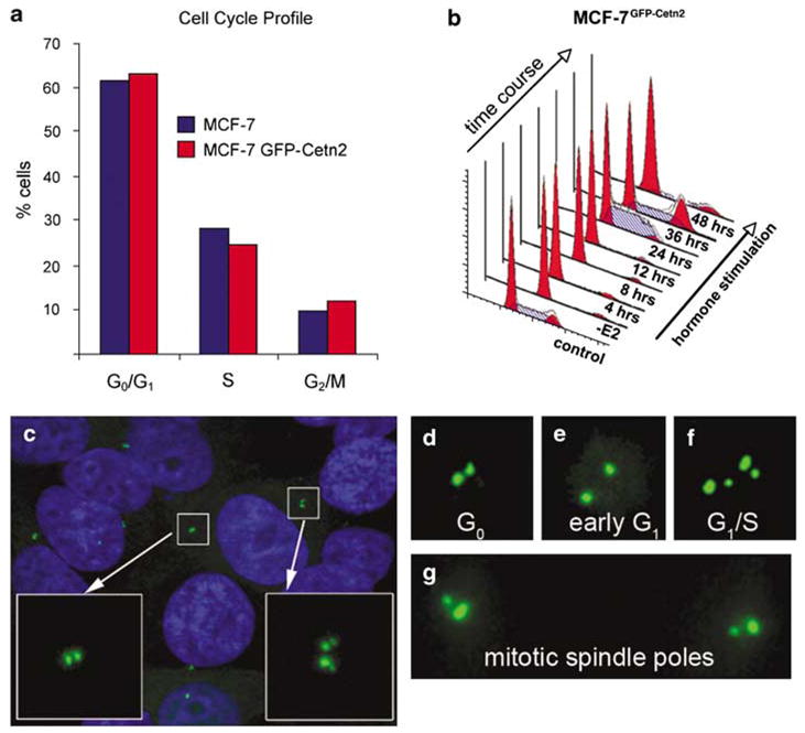
Centriole dynamics during cell cycle progression in MCF-7 cells. (a) Cell cycle analysis (fluorescence-activated cell sorting) for MCF-7 and MCF-7GFP–cetn2 cells showing that green fluorescent protein(GFP)-centrin does not alter cell cycle kinetics. (b) Cell cycle progression following mitogen stimulation in the MCF-7GFP–cetn2 cells. –E2: 48-h withdrawal of 17-β estradiol, EGF and IGF-I. (c) MCF-7GFP–cetn2 cells showing different centriole duplication stages. GFP-centrin (green) and Hoechst stained nuclei (blue). (d) G0 phase with a pair of centrioles adjacent to one another. (e) Early G1 phase with centrioles separated from one another. (f) Late G1 or early S phase characterized by centriole duplication. (g) G2/M phase with a pair of centrioles at each mitotic spindle pole.