
Figure 3.
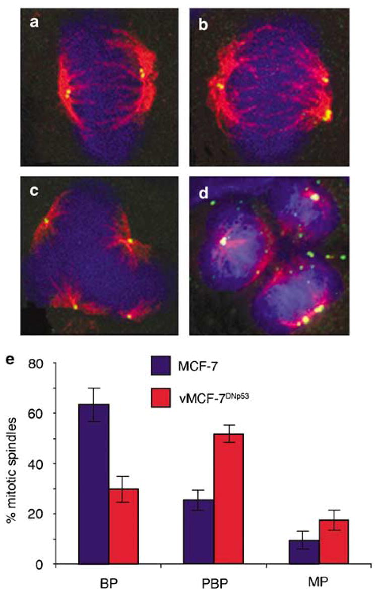
Mitotic spindle morphology in MCF-7 and vMCF-7DNP53 cells. (a–d) Immunofluorescence of bipolar (a) pseudobipolar (b) and multipolar mitotic spindles (c) in the vMCF-7DNP53 cells. (d) An example of a multipolar mitosis resulting in the generation of more than two daughter cells. Centrioles were labeled for centrin (green), mitotic spindles were labeled for the centrosome kinase aurora-A (red) and DNA with Hoechst dye (blue). (e) Graph showing the percentage of mitotic spindles from three experiments±s.d. with bipolar (BP), pseudobipolar (PBP) and multipolar (MP) morphology cells following 24-h mitogen stimulation.