
Figure 4.
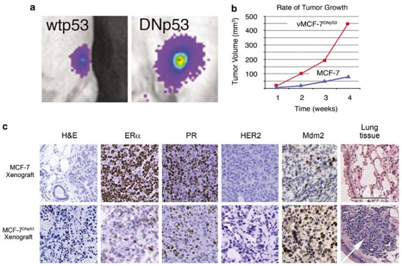
Human breast cancer xenografts in nude mice. (a) Tumor imaging in live animals of MCF-7 (left) and vMCF-7DNp53 (right) xenografts expressing the firefly luciferase reporter 4 weeks after subcutaneous injection. (b) Measurement of weekly tumor growth using digital calipers. (c) Paraffin sections of xenograft tumors (12 weeks) showing: H&E staining of low-grade tubular tumors for MCF-7 and anaplastic vMCF-7DNp53 tumors; loss of estrogen receptor α and progesterone receptor expression in the vMCF-7DNp53 xenografts; negative phenotype for Her2/nue in both xenografts; elevated MDM2 staining and metastatic spread to lungs for the vMCF-7DNp53 xenograft.