
Figure 5.
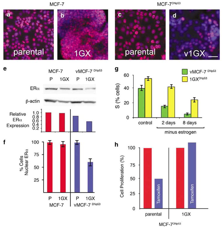
Estrogen receptorα status, estrogen-dependent growth, and tamoxifen sensitivity for MCF-7 and vMCF-7DNp53 and cultures reestablished (1GX) from xenograft tumors. (a–d) ERα staining (red) showing ERα expression and nuclear localization in MCF-7 (a), vMCF-7DNp53 (c), and recultured 1GX MCF-7 (b). Loss of expression and nuclear localization was seen only in the recultured v1GX MCF-7DNp53 cells (d). Nuclei were counterstained for DNA using Hoechst dye (blue). (e) Western analysis of ERα abundance showing reduced expression in the 1GX vMCF-7DNp53 cells. ERα expression relative to β-actin the loading control quantified by densitometry. (f) Quantitative analysis of cells with ERα nuclear localization. (g) FACS analysis of % cells in S-phase before and following estrogen withdrawal showing reduced hormone dependence in v1GX MCF-7DNp53 compared to the parental vMCF-7DNp53 cells. (h) Cell proliferation assay showing loss of tamoxifen sensitivity in the cultures reestablished from v1GX MCF-7DNp53 xenografts.