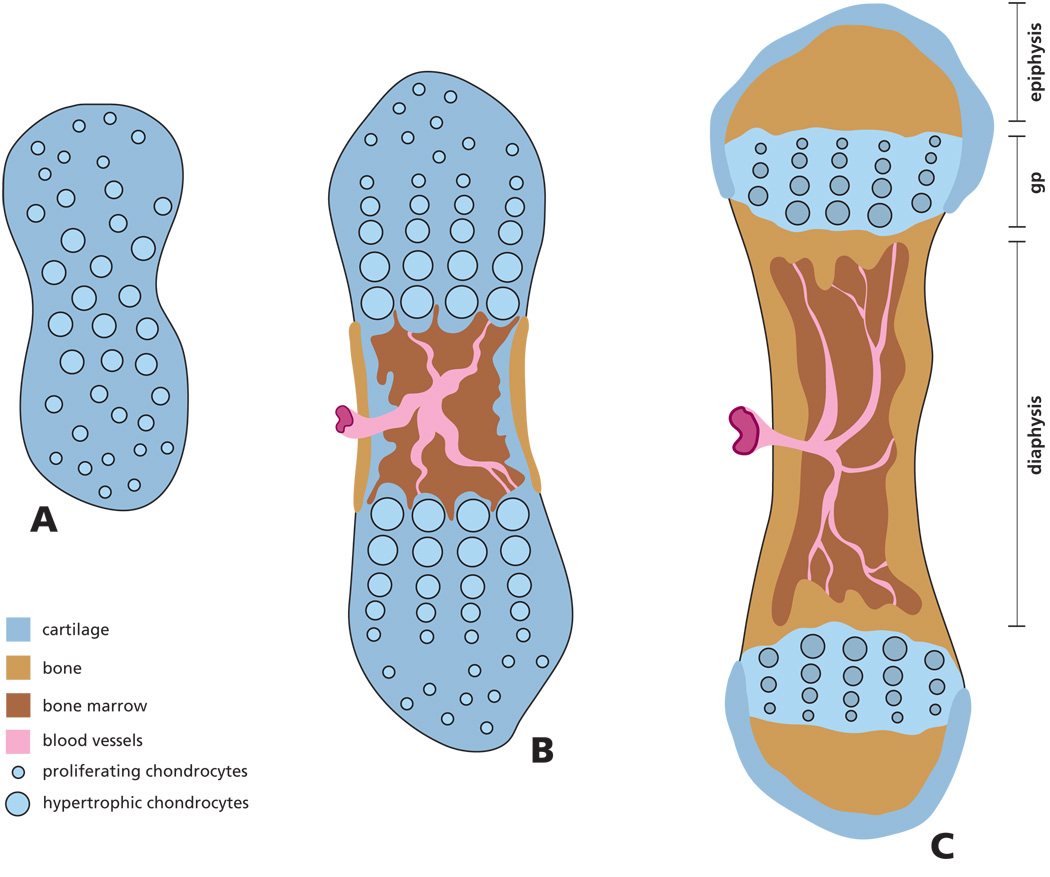
Figure 1. Scheme of endochondral bone formation during development.
Initially the mesenchyme in the region of the future bone anlage differentiates into chondrocytes, forming a cartilage model of the future bone (A). Cells in the center of the cartilage model undergo further differentiation into hypertrophic cells inducing vascular invasion of the diaphysis and formation of a marrow cavity. As a result of osteoclast and osteoblast activity, the cartilage is resorbed and the first osteoid is deposited (B). Areas of secondary ossification develop in the epiphysis, while in the growth plate (gp), located between the epiphysis and diaphysis, columns of chondrocytes continue to proliferate, hypertrophy, secrete and mineralize the extracellular matrix (C). Over time this matrix is partially resorbed and replaced with new bone resulting in bone elongation.

