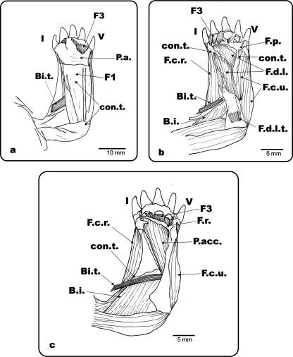
Fig. 4.
Ge. chilensis. Ventral view of the left antebrachium and hand. (a) Superficial musculature and connective tissue of the antebrachium and hand; (b) palmar aponeurosis, Fusion 1 and most of the superficial connective tissue removed; (c) flexor digitorum longus and flexor plate removed. F.d.l.: m. flexor digitorum longus; F.d.l.t.: m. flexor digitorum longus tendon; P.a.: palmar aponeurosis; con.t.: connective tissue; F.p.: flexor plate; F.c.r.: m. flexor carpi radialis; F.c.u.: m. flexor carpi ulnaris; Bi.: m. biceps; Bi.t.: m. biceps tendon; P.t.: m. pronator teres; B.i.: m. brachialis inferior; P.acc.: m. pronator accesorius; F.r.: flexor retinaculum; F1: fusion 1; F3: fusion 3; I and V: digits I and V.

