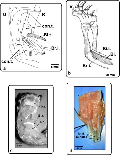
Fig. 5.
(a) Ge. chilensis, ventral view of the right antebrachium and hand. (b) Ph. hilarii, ventral view of the left antebrachium and hand. (c) Ge. chilensis, photograph of the biceps tendon insertion. A ventral view of the right antebrachium and hand (d) Muscle extensor digitorum longus removed from the dorsal side of the forelimb. Colour photograph showing the ventral face of the muscle with the tendinous bundles of its head. Bi.: m. biceps; Bi.t.: m. biceps tendon; Br.i.: m. brachialis inferior; R: radius; U: ulna; con.t.: connective tissue; P.a.: palmar aponeurosis; Tend. Bundles: tendinous bundles; I and V: digits I and V.

