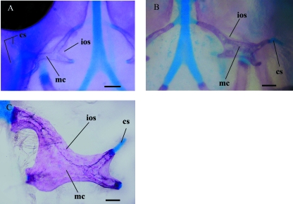
Fig. 5.
Early orbitosphenoid development showing the compound nature of the bone from cleared and double-stained material. (A) Left orbitosphenoid of embryo I in dorsal view. (B) Right orbitosphenoid of embryo II in dorsal view. (C) Right orbitosphenoid of a neonate (UIS-R-1452) in dorsal view. cs, cartilaginous scaffolding; ios, intramembranous ossification; mc, medullar cavity. Scale bars = 100 µm.

