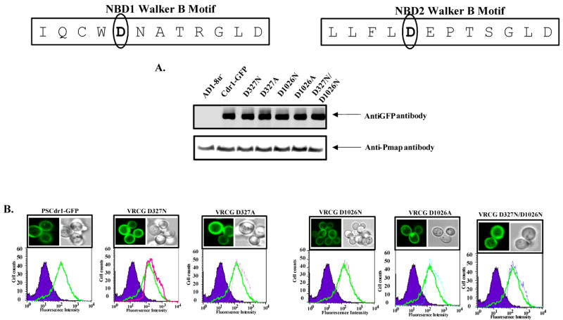
Figure 2. Membrane localization and expression profile of wild type and carboxylate residue mutant variant Cdr1p.

Boxed panel at the top shows the sequence of Walker B and extended Walker B motifs of N-terminal and C-terminal NBDs of Cdr1p. (A) PM of wild type and mutant variant proteins expressing cells were prepared and their immunodetection was done as described earlier (26).
(B) Fluorescence imaging (upper panel) by a confocal microscope showing membrane localization of Cdr1-GFP (Cdr1p) and its mutant variant proteins expressing cells. Flow cytometry (lower panel) of S. cerevisiae expressing Cdr1p and its mutant variants. The histogram derived from the cell quest program depicts fluorescence intensities for AD1-8u- (control) (purple filled area), PSCdr1-GFP (solid green line) for each panel and other extra line represent that respective Cdr1p mutant variant expressing cells.
