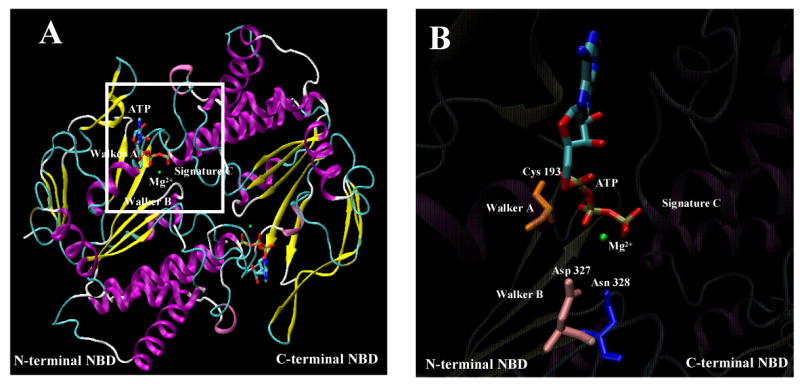
Figure 7. Atomic model of the N-and- C-terminal NBD dimer of Cdr1p.

(A) Homology based model of N-and-C-terminal NBD of Cdr1p. The structure of N- and C-terminal NBD was generated by Modeller8v2 program (19,31). Structural diagrams were produced using Visual Molecular Dynamic (VMD) software (29), where α-helices are represented as ribbons and strands as arrows. The ATP molecule is shown in licorice format and the atoms, i.e., carbon, nitrogen, oxygen, phosphorus, sulfur, and magnesium ion, are represented in cyan, blue, red, tan, yellow, and green, respectively. Functionally important sequence motifs of NBDs are labeled. (B) A blown-up view of the N-terminal nucleotide binding pocket of the dimeric model of Cdr1p highlighting the close positioning of ATP and Mg2+ with Asp327 (pink), Asn328 (blue), Trp326 (green) and Cys193 (orange) residue side chain.
