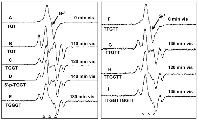
Figure 3.
ESR spectra of (A) and (F) of one-electron oxidized TGT (4.5 mg/ml) and TTGTT (4.5 mg/ml) formed by attack of Cl2•– in the presence K2S2O8 (8 mg/ml) as an electron scavenger in 7.5 M LiCl glass / D2O, in annealing to 155 K. These spectra in (A) and (F) are chiefly due to G•+. Spectra (B) – (E) and (G) – (I) are obtained after photo-excitation of G•+ in the oligonucleotides indicated with visible light at 143 K, showing considerable conversion of G•+ to sugar radicals - C5′• (prominent doublet at the centre) as well as to C1′• (prominent quartet). The samples in A and (F) are red-violet due to the absorption of G•+ whereas those in (B) – (E) and (G) – (I) are fade in color or are near colorless as conversion to sugar radical occurs. All spectra were recorded at 77 K.

